
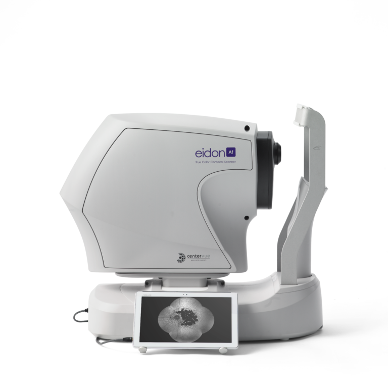
iCare EIDON AF
iCare EIDON AF blue autofluorescence confocal fundus imaging system
Key features
- TrueColor, blue autofluorescence, infrared and RGB channels confocal images
- Panoramic view of the retinal autofluorescence (up to 160°)
- High details and contrast in the same device
- Short exam time and enhanced patient comfort
- Easy to use, speeds up patient workflow
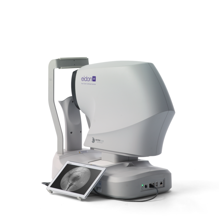
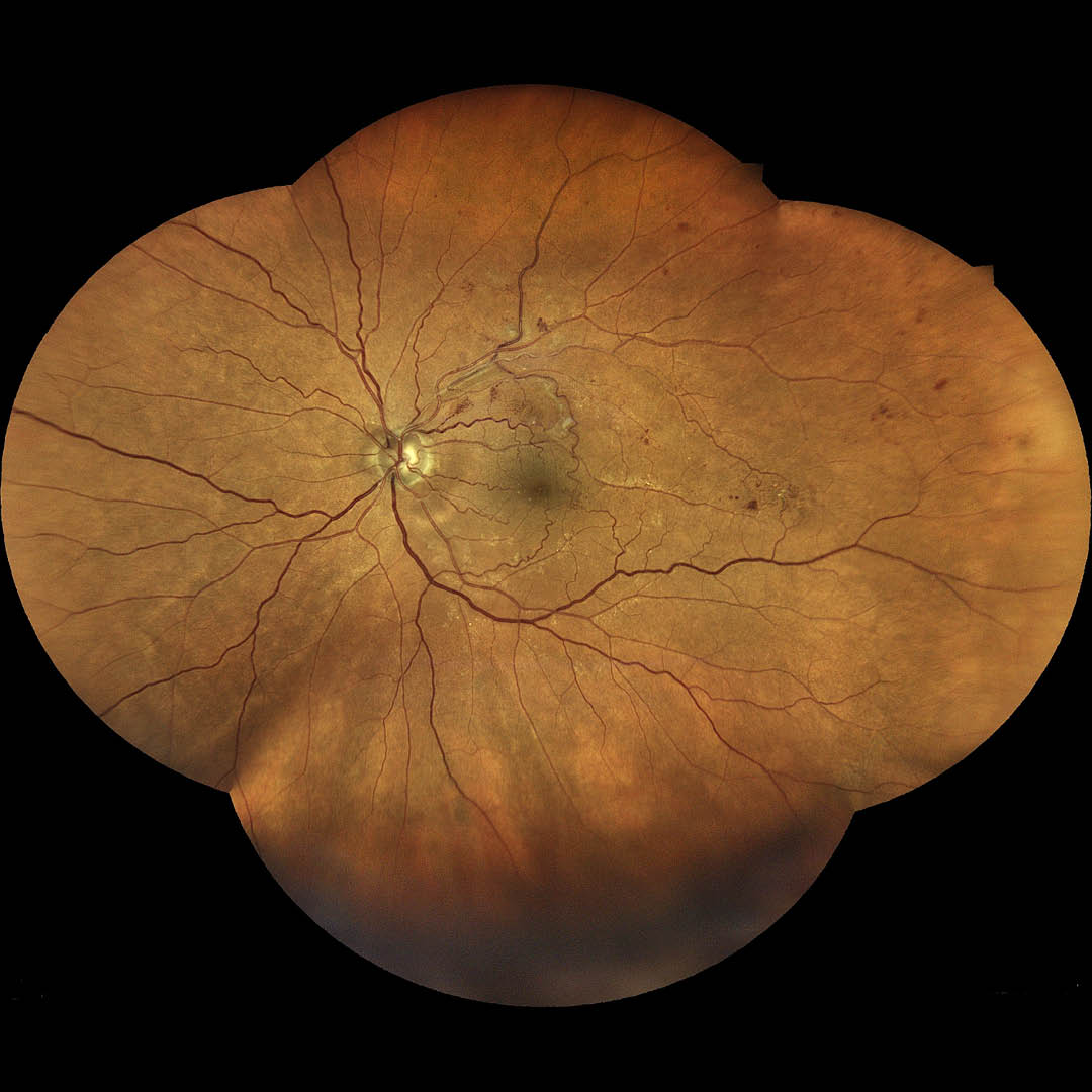
iCare EIDON AF TrueColor high-resolution confocal fundus retinal imaging system with blue autofluorescence (FAF)
iCare EIDON AF TrueColor confocal scanner offers the best of iCare EIDON technology with the added advantage of autofluorescence imaging capabilities. The fully automated scanner captures high resolution, accurate imaging using multiple modalities — TrueColor, red and blue imaging, red-free, infrared and blue autofluorescence. The cutting-edge device obtains excellent images in pupils as small as 2.5 mm.
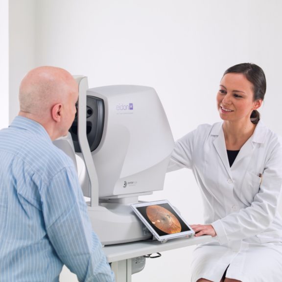
The quick and non-invasive autofluorescence imaging technique improves patient comfort and experience. With a fully computer-assisted acquisition mode, any staff member can run tests, which can help reduce waiting time and aid in improving workflow efficiency. This device truly offers the best to both patients and eyecare professionals.
Multiple modalities for a complete picture
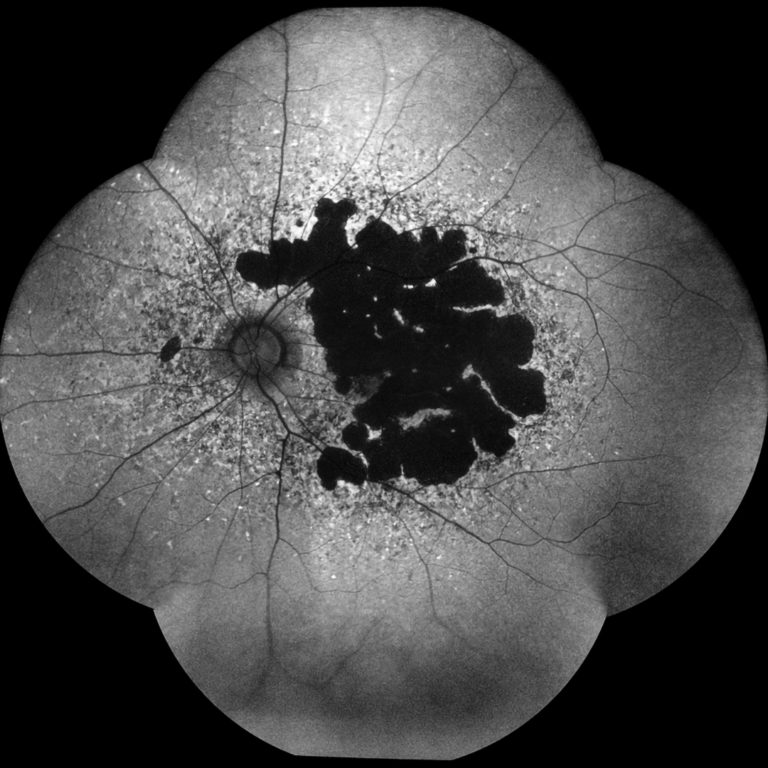
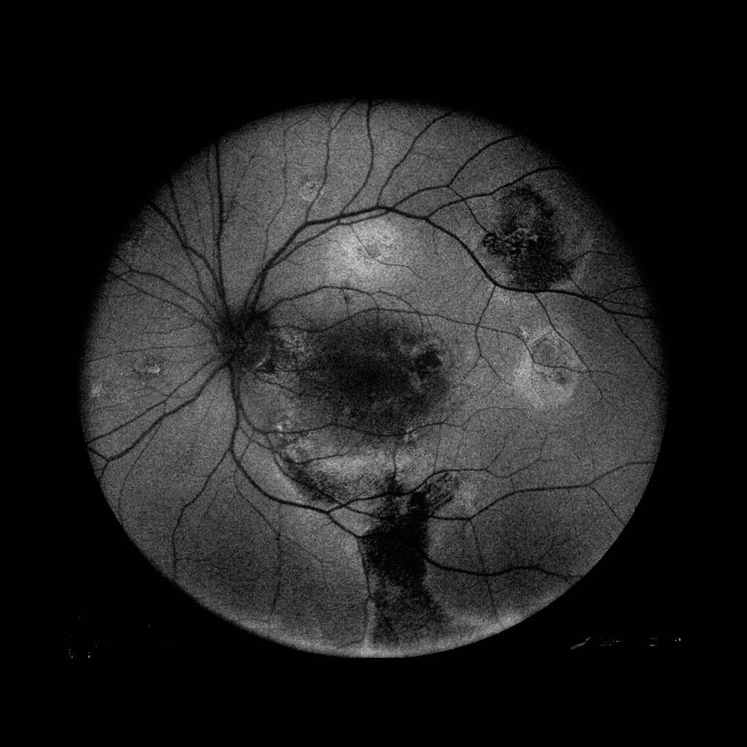
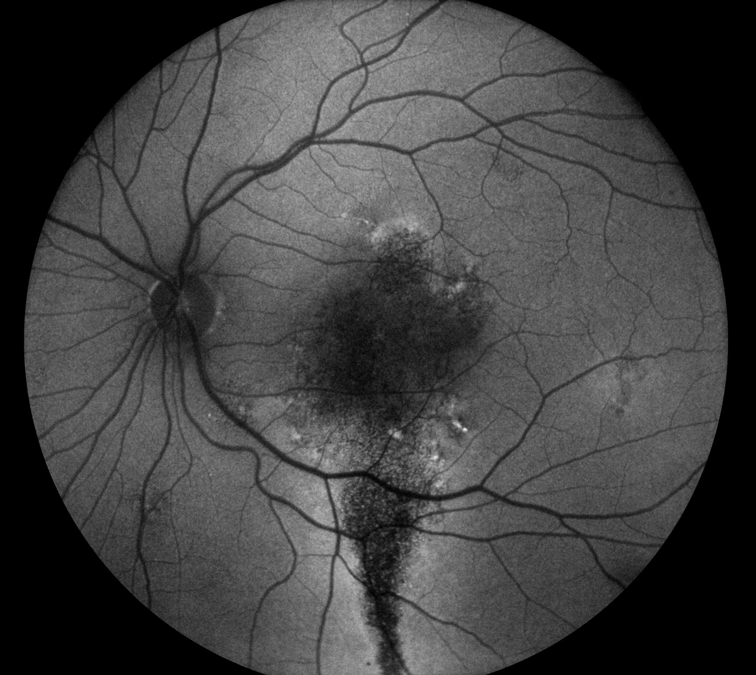
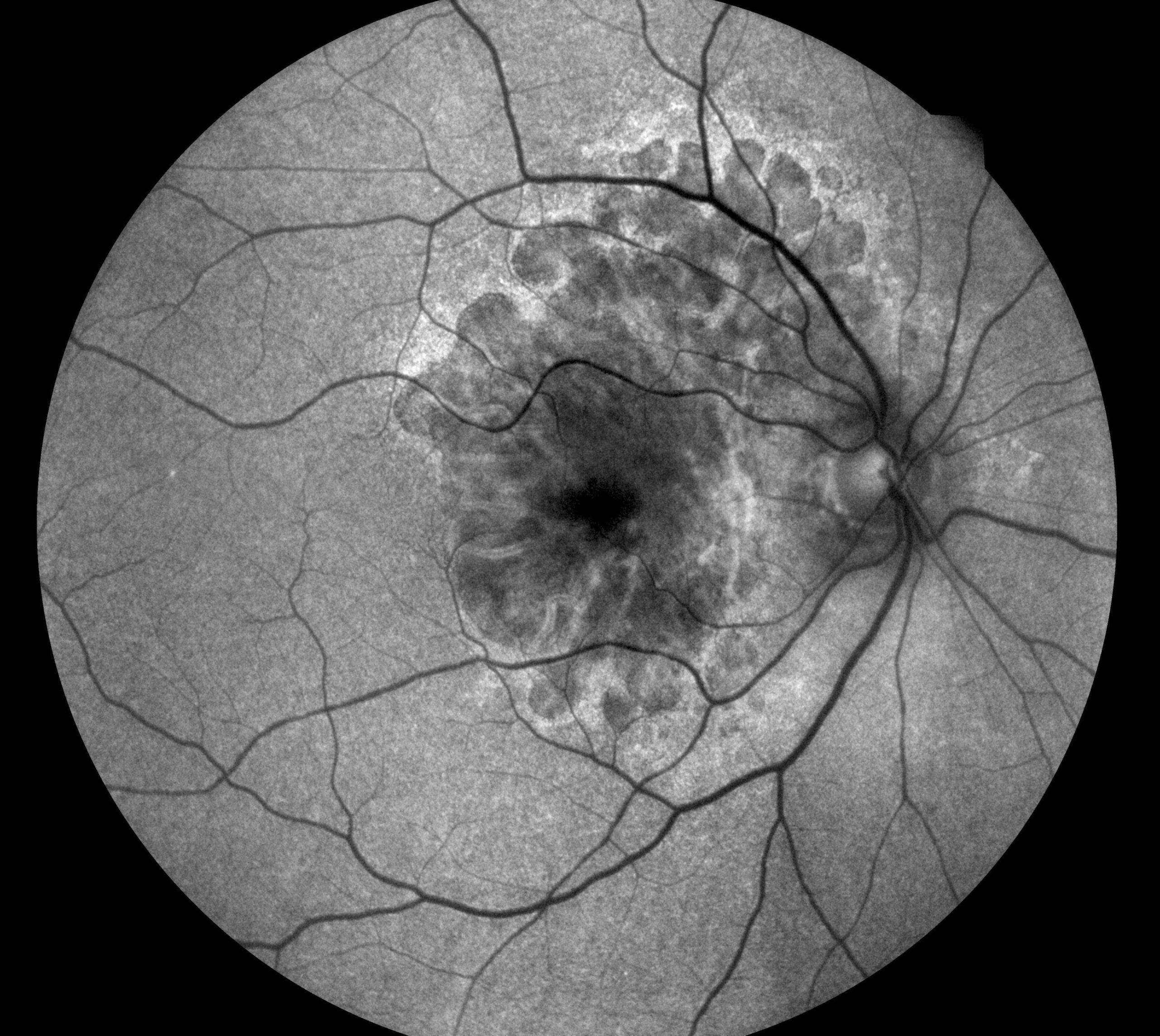
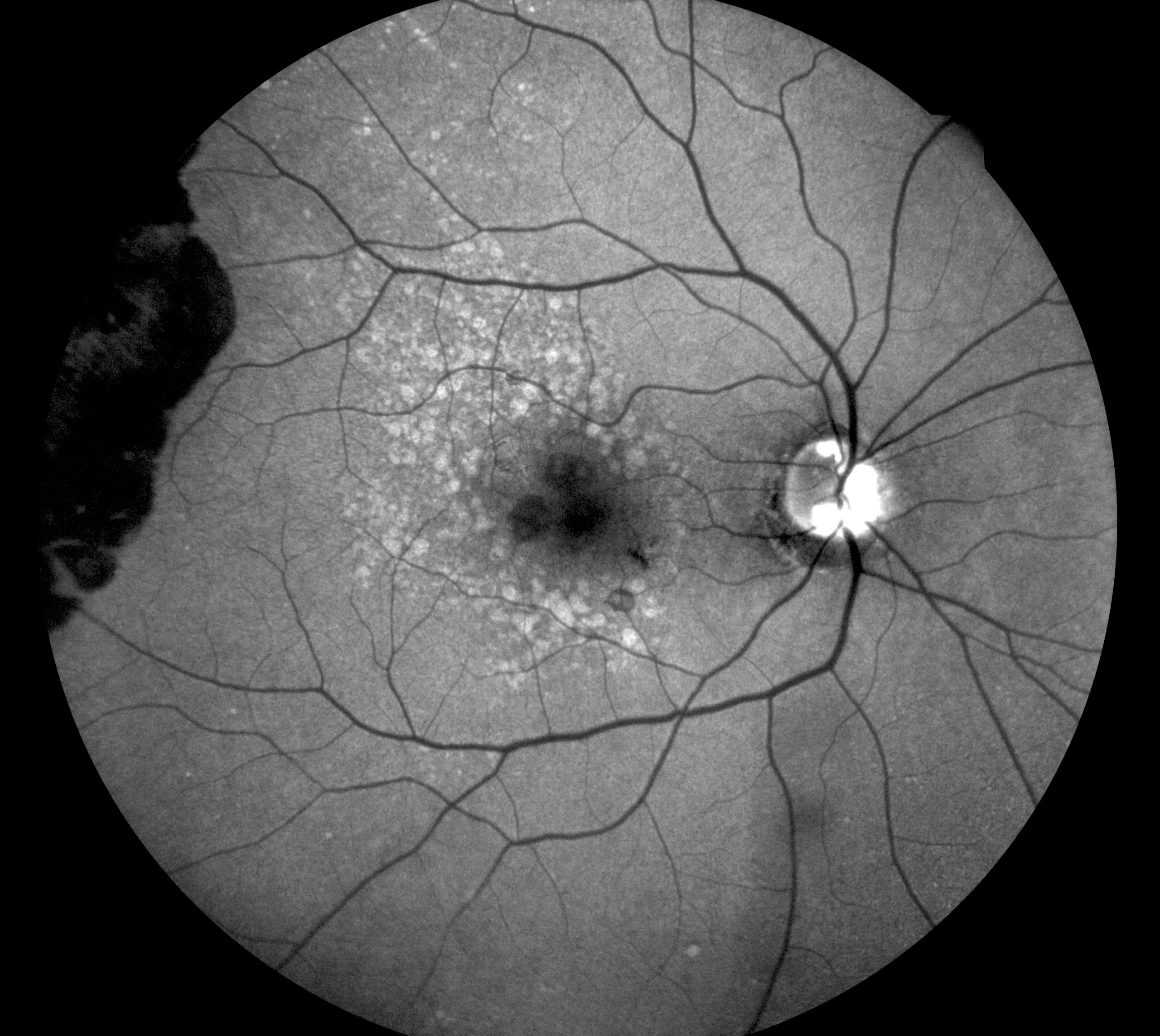
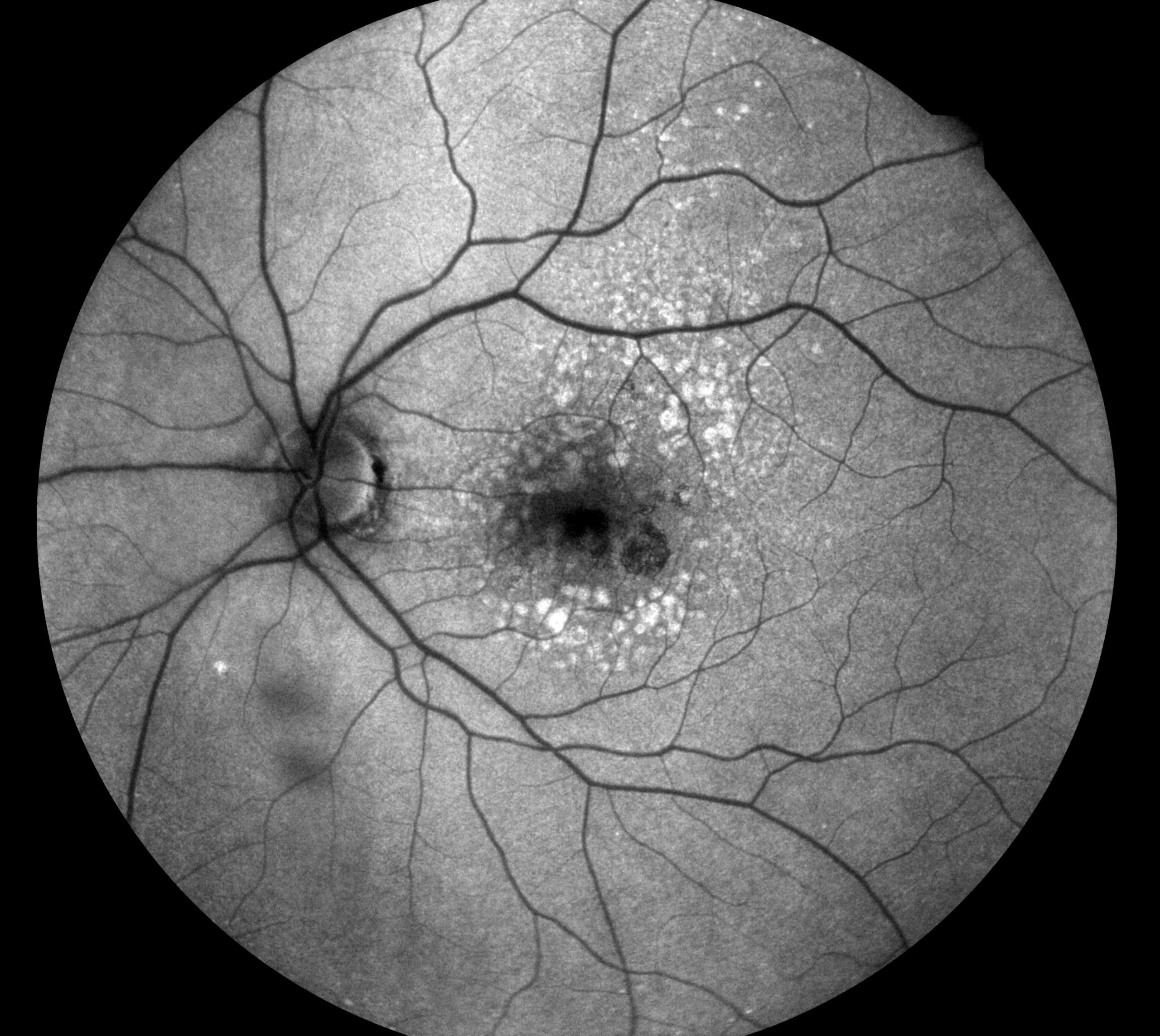
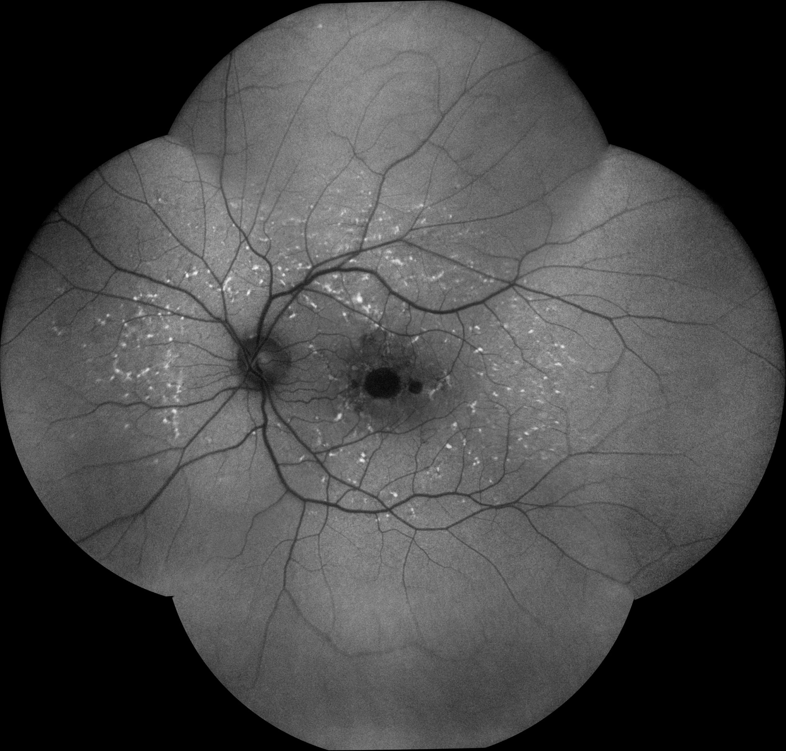
iCare EIDON AF obtains multiple types of high value information from multiple imaging modalities to help determine an accurate diagnosis. White LED illumination provides high-quality TrueColor images, Red-free filtering enhances visualization of retina vasculature, blue images provide improved view of the Retinal Nerve Fiber Layer (RNFL) and red channel allows to penetrate in the deeper layers towards the choroid. Infrared light provides detailed information corresponding to the choroid. Autofluorescence allows the assessment of the Retinal Pigment Epithelial (RPE) layer integrity.
The advantage of wide field of view acquisition capability
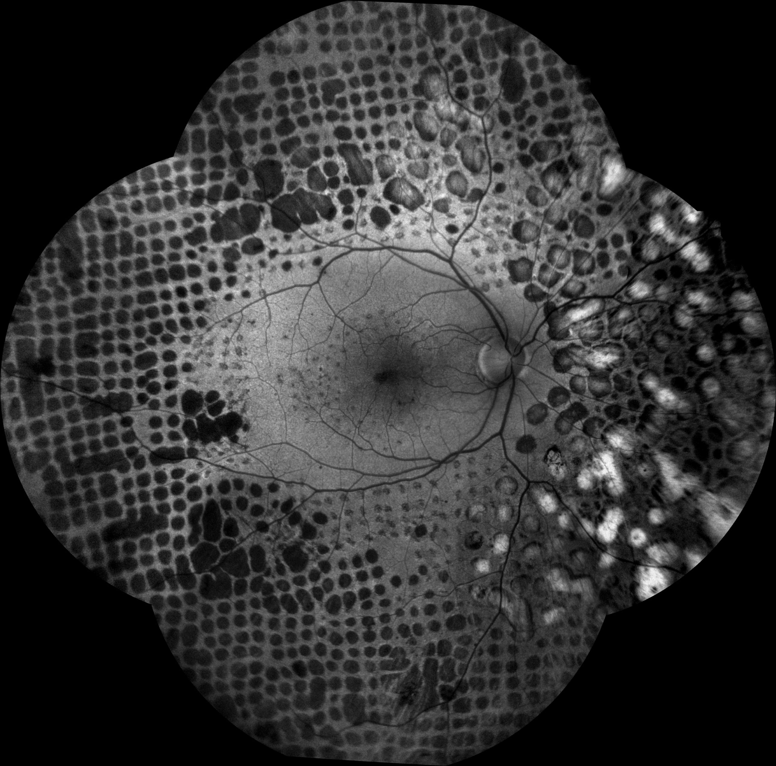
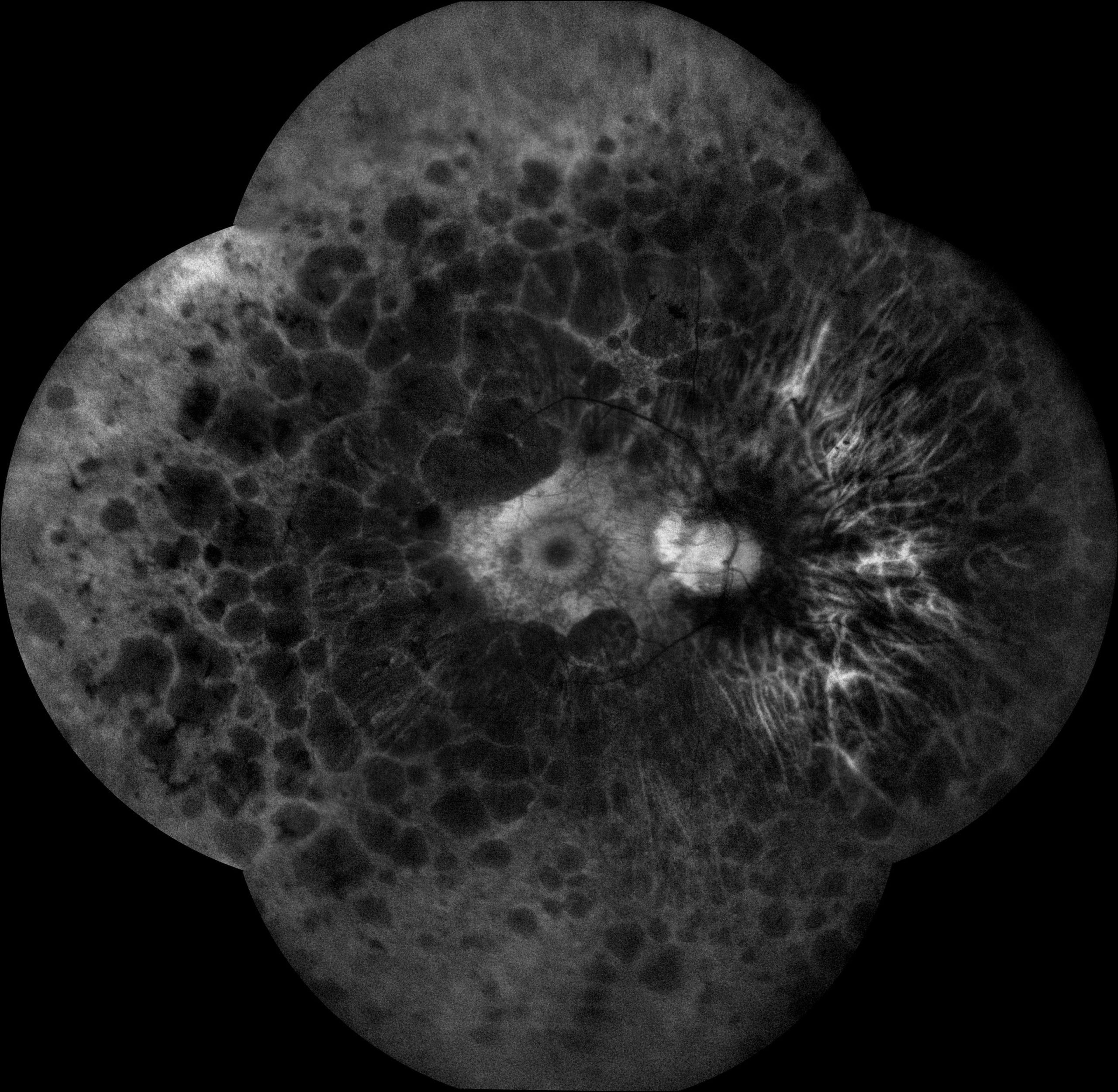
iCare EIDON AF offers the added advantage of capturing high quality Widefield images, up to 160°, thanks to its unique mosaic functionality. This capability, available for all the different imaging modalities (TrueColor, IR, FAF) plays a crucial part in supporting eyecare professionals discover, diagnose, document and treat ocular pathologies that may first present in the periphery.
Increased field of view up to 200˚
Thanks to the EIDON Ultra-Widefield Module it is possible to increase the field of view up to 200˚, which helps to detect signs of pathologies that start to appear in the periphery. The Ultra-Widefield module enables retina from 120° with a single shot, up to 200° with Mosaic functionality.
Unsurpassed image quality in retinal imaging
The confocal scanner guarantees high details and contrast Autofluorescence images with an ultra-high resolution quality in one shot, without the need of image averaging — this makes for a quick and comfortable exam experience for the patient. Furthermore, the confocal technology allows scanning through cataract and other media opacities. It also offers 60° autofluorescence images with a single flash of light and a panoramic view (up to 110°) of the retinal autofluorescence.
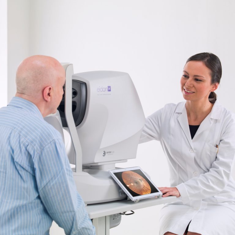
Fast and fully automated retinal imaging process
In the automatic mode, the scanner auto-aligns to a patient’s pupil, focuses on the retina and captures high-quality, accurate images in any of the imaging modalities available: IR and/or color and/or FAF, allowing any staff member to run tests effortlessly. The manual mode offers improved versatility — it allows one to customize the management of the focus, concentrate on specific layers of the fundus and go wider in periphery using the external fixation target. The easy-to-use and compact imaging device speeds up patient workflow efficiently.
Patient comfort and convenience
The quick examination time makes it an easy and comfortable experience for patients. The non-invasive Fundus Autofluorescence (FAF) imaging technique used in the iCare EIDON AF retinal imager uses a single flash of light which ensures maximum patient comfort. The images can be captured even in a pupil as small as 2.5 mm, making it possible to obtain high-quality images without dilation.
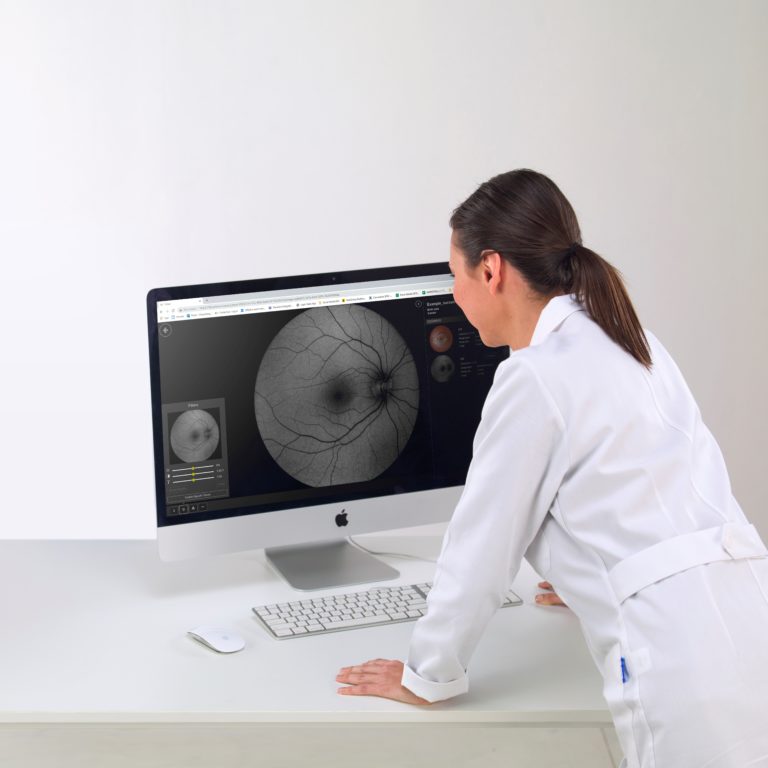
Image sharing and viewing made easy
EIDON AF multi-touch tablet makes magnification and review easy. Additionally, EIDON AF Remote Viewer (RV) makes remote data review effortless, allowing any computer or laptop on the same local area network (LAN) to review EIDON AF images remotely, enabling data access and detailed analysis on multiple review stations.